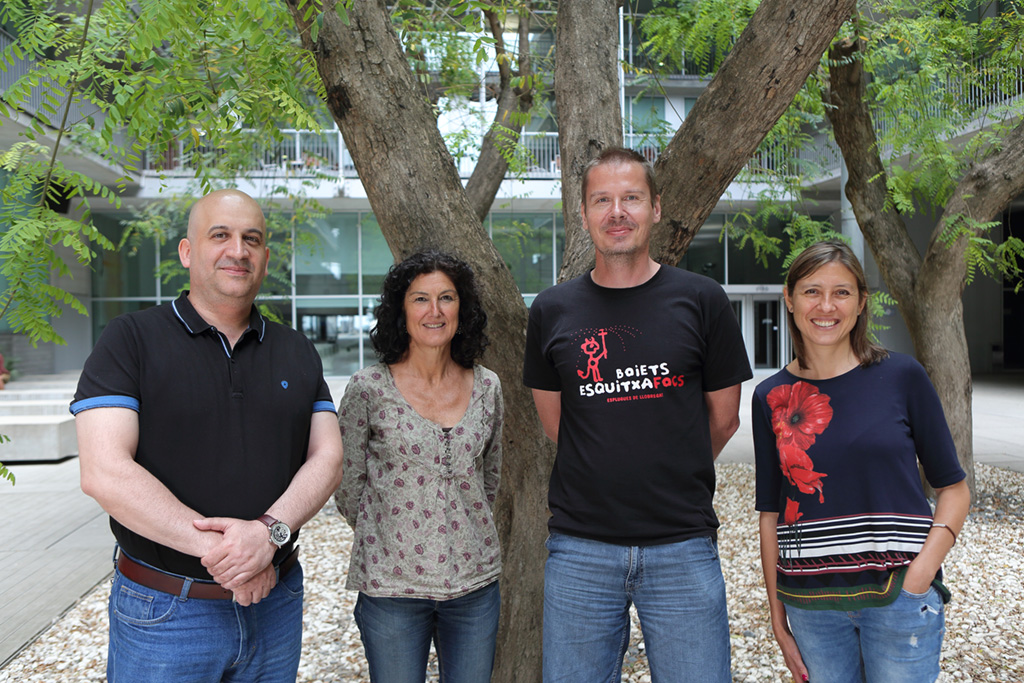
HEAD OF FACILITY:
Timo Zimmermann
MICROSCOPY SPECIALISTS:
Raquel Garcia Olivas, Arrate Mallabiabarrena, Xavier Sanjuan (UPF)
SCREENING AND AUTOMATION:
Raul Gomez
HEAD OF FACILITY:
Timo Zimmermann
MICROSCOPY SPECIALISTS:
Raquel Garcia Olivas, Arrate Mallabiabarrena, Xavier Sanjuan (UPF)
SCREENING AND AUTOMATION:
Raul Gomez
The Advanced Light Microscopy Unit (ALMU) of the CRG and UPF serves as a core facility for high-end light microscopy for PRBB researchers. A range of instruments with unique capabilities fully covers the spectrum of advanced imaging applications from thick tissue reconstruction to fast in-vivo imaging to the sensitive detection of very faint signals of single molecules. The staff of the facility provides advice in the initial experiment planning, training of the researchers on the instruments and assistance with the subsequent data analysis. It is the aim of the facility to provide a link for the biological questions of researchers to the full capabilities of advanced light microscopy at the organismic, cellular and molecular level. Methods available in the facility include super-resolution microscopy by stimulated emission depletion (STED) and localization based methods like Stochastic Optical Reconstruction Microscopy (STORM) and Ground State Depletion Imaging Microscopy (GSDIM), optical sectioning (single photon and multi-photon microscopy), spectral imaging, in-vivo time-lapse imaging, Total Internal Reflection Fluorescence (TIRF) Microscopy and methods for the study of molecular properties and interactions like Fluorescence Correlation Spectroscopy (FCS), Fluorescence Lifetime Imaging Microscopy (FLIM), Fluorescence Resonance Energy Transfer (FRET) detection, Fluorescence Recovery after Photobleaching (FRAP) and confocal and widefield screening microscopy. Additionally, dedicated software packages for data visualization and analysis are available for 3D rendering, particle tracking and image analysis.
In 2017, the total booked microscope usage time of the unit reached more than 18000 hours in more than 6000 separate bookings. This corresponds to approximately seven hours of daily usage on the bookable microscope systems plus many additional hours on equipment without mandatory booking and on special equipment. During the year, 123 users from 29 CRG research groups and 59 users from 22 UPF-CEXS groups have used the unit. Additionally, the unit was used by 34 users from 16 groups of other PRBB institutes and one company and for projects from external visitors. Towards the end of the year, the unit also started to provide microscopy service to the newly created EMBL Outstation. On average, 92 investigators use the unit every month.
After several months of technical refurbishments and general renovation of the area, the ALMU started towards the end of 2017 to move into its new location in the basement of the PRBB building. This on the one hand means an increased physical distance from the CRG research groups (and losing one of the most spectacular locations in the world for a microscopy unit on the fifth floor that we truly enjoyed over the last ten years). But this is offset by expanding into two main instrument rooms that allow us to better separate and maintain the advanced microscopy systems as the upstairs location couldn’t suitably accommodate the number of systems anymore. It also provides additional experiment preparation space and a microscope development room. Most importantly, the newly created technical space will be shared with the neighbouring installations of the newly established imaging unit of the EMBL Barcelona Outstation for Tissue Biology and Disease Modelling. This opens exciting possibilities of synergies between complementary imaging fields in a total space of more than 330 sqm (distributed evenly between the units) that will be exclusively dedicated to high-end imaging methods and image-based analysis and modelling.
The technology offer of the unit was kept up-to-date by the following additions:
Timo Zimmermann is the Spanish representative for Biological Imaging in the Interim Board of the ESFRI project Euro-BioImaging. The microscopy unit participates in two highly evaluated Spanish node proposals for Euro-BioImaging, for super-resolution microscopy (with the Photonic Sciences Institute ICFO) and for in-vivo and intravital imaging (with the University of Barcelona and IRB Barcelona) which are participating in the interim operation phase of Euro-BioImaging which has started in May 2016.
All members of the staff are frequently participating as speakers and instructors in master’s courses from the CRG and UPF, as well as in many conferences and microscopy courses both at the PRBB as well as at other institutions, nationally and internationally.
The unit participated in the execution of the Summer School of Molecular and Theoretical Biology organized for Russian high school students at the CRG and UPF.